
|
Register |
Take a Quiz |
Leave Feedback - New Window |
Consult Request Form (PDF)
|
|
Highlight from this week
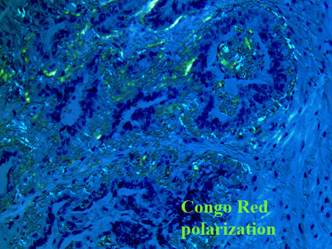
Cross-sections of the seminal vesicles are free of tumor, but show dense subepithelial deposits of an amorphous hyaline material. Congo red stain shows apple green birefringence. There is prominent amyloid deposition within the stroma of the seminal vesicles and vasa deferentia, with no vascular amyloid deposition identified. |
This on-line interactive, CME activity is sponsored by the University of Pittsburgh School of Medicine, Center for Continuing Education in the Health Sciences and the Department of Pathology.
The target audience for this activity is pathologists and other physicians, especially those practicing in remote and/or underserved areas. At the end of the presentations, the participants should be able to:
The University of Pittsburgh School of Medicine, as part of the Consortium for Academic Continuing Medical Education, is accredited by the Accreditation Council for Continuing Medical Education to provide continuing medical education for physicians. The Center for Continuing Education in the Health Sciences designates this educational activity for up to a maximum of 1.0 hour of Category 1 credit towards the AMA Physician's Recognition Award. Each physician should claim only those hours of credit that he/she actually spent in the educational activity.
The authors for each case are listed on header of the appropriate cases. The pathology faculty from the University of Pittsburgh responsible for the site and for editing and reviewing cases include:
Neither The University of Pittsburgh School of Medicine, Center for Continuing Education in the Health Sciences nor the UPMC Department of Pathology, will sell, trade, rent or share personal information to any commercial or external entity. The information collected will be used for managing and monitoring continuing medical education credits and evaluation purposes.